


")


.png "Interview With Expat Saanika Gandhi, Author of Children's Book 'The Yellow Kite'")


Your eyeballs' anatomies are actually made up of loads of amazing things that help give you your sight
31 January 2017
| Last updated on 5 February 2017
Do You Actually Know What's In Your Eye?
While small in size, our eyes give us one of our important senses... Vision.
Some people call them the window to the soul, but while they may be dazzling to look at, they're a lot more complicated than what you might think.
An eye is simply not just an eye. Both of yours are actually made up of different parts to create its anatomy. In fact, there's 13 different parts that make up a human eye, and here we take a peek (pun intended) into each one...


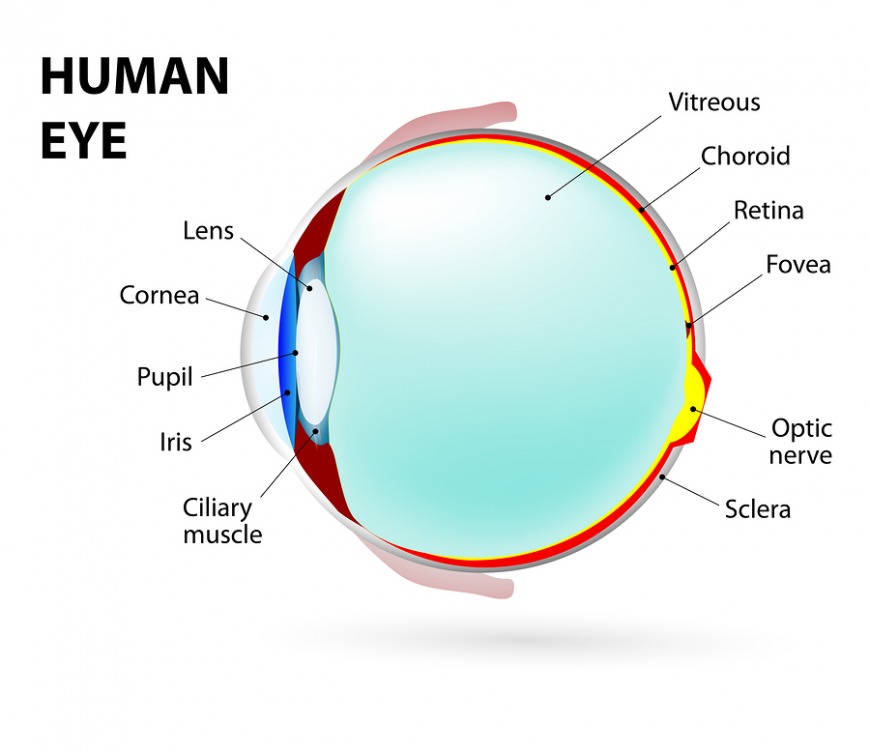

The cornea
This is the transparent circular part of the front of your eyeball. It refracts the light entering into the eye onto the lens, which then focuses it onto the retina. Your corneas are the only parts of your body that do not have blood vessels, and they're extremely sensitive to pain.
The iris
This regulates the amount of light that enters your eyes. It forms the coloured part of your eye, and light enters them through the pupil.
The pupil
Behind your cornea is the circular opening in the centre of the iris, which light passes through and into the lens of the eye. Your iris controls the widening and narrowing (dilation and constriction) of your pupil depending your environment.
You might also be interested in:
- 5 simple eye care tips for computer users
- How air conditioning can ruin your eyesight
- 13 fascinating facts about the human eye
The lens
This is a transparent structure found behind the pupil of your eye, and it's in a thin, transparent capsule. It helps to refract incoming light and then focuses it onto your retina. Did you know? When people have cataracts, this means their lens has become cloudy and to fix this in a cataract operation means replacing the cloudy lens with a plastic one.
The ciliary body
This is the part of the eye that connects the choroid to the iris.
The choroid
This is the middle layer between your eye, retina and the sclera. It's useful as it contains a pigment that absorbs excess light, which helps to prevent blurry vision.
The retina
Your retinas are a sensitive layer that lines the interior of your eyes. They're made up of light sensitive cells called rods and cones. In one human eye, there's around 125 million rods, which are necessary for seeing in dim light. On the flip side, cones function best in bright light and there's between 6 and 7 million of these in one eye. They're essential so you can see a sharp accurate image and distinguish colour.

The sclera
This is the white part of our eyes, and is a tough covering with which the cornea forms the external protective coat of the eye.
The optic disk
This is visible when your eye is examined, and is found on the retina of your eye. The optic disk identifies the start of the optic nerve, where messages from your cone and rod cells leave the eye by nerve fibres and send them to the brain. This area is actually a blind spot in your eyes.
SEE ALSO: What really happens when you sleep in your contact lenses
The optic nerve
This leaves your eye at the optic disk and transfers all visual information to your brain.
The fovea
This is a small identation at the centre of the macula, and is described as an area with the greatest concentration of cone cells.
The macula
This is a yellow spot on your retina at the back of the eye, which surrounds the fovea. There's a great concentration of cone cells here and when your eye sees something, the part of the image that is focused on the fovea is the image most accurately registered by the brain.
The vitreous
This is a clear gel that fills the space between the lens and the retina of our eyeball. Often referred to as the vitreous humour.


")


